Description
Excellent cardiovascular performance
Tissue Doppler Imaging (TDI)
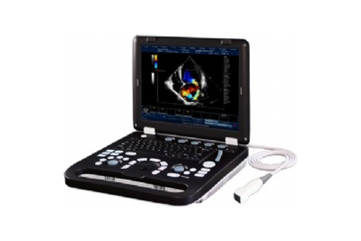
- As a noble echocardiography technique, Tissue Doppler Imaging is used for myocardial velocity measurement. Systolic TD measurements are used for evaluation of left and right ventricular myocardial contraction functions, while diastolic TD values reflect myocardial relaxation.
Auto IMT
- Dedicated software measures carotid Intima Media Thickness(IMT) automatically and accurately.
Free Steer M
- Anatomical M mode with three sampling lines shows motions of three cardiac regions simultaneously, which greatly improves diagnostic efficiency.
- Monitor: 15inch LED
- Probe connector: 1 (option 2/3)
- TGC: 8 segments
- Gray scale: 256
- Bodymark: 123 types
- Scanning method: electronic convex / linear / micro-convex / phased-array
- Supporting probe: electronic convex / linear, broadband, multi-frequency, THI on all probes
- Image processing: image enhancement, speckle reduction, rejection, lines density
- Clinical application: abdominal, obstetric, gynecology, cardiology, urology, small part, etc
- Image mode: B, 2B, 4B, B/M, CFM, B/BC, Color M, PW, CW, PDI, DPDI, Duplex, Triplex,
- Panoramic, Trapezoid
- Focus depth: 0-10, 5-16, 10-35
- Focus numbers/set: 1, 2, 3, 4
- Frame average: 0(N/A), 2, 3, 4, 5, 6, 7, 8
- Dynamic range: 20-80 dB, 6-scale interval adjustable
- Lines density: low, medium, standard, high
- Rejection: 0-32
- Speckle reduction: 1-8 levels NeatView / PureView
- Image enhancement: 1, 2, 3, 4 method
- Scanning angle: 40 to 160 degrees, depending on probe
- Scanning depth: 30-300mm, depending on probe
- View area: 50 / 60 / 70 / 80 / 90 / 100%
- Image flip: up / down, left / right, black / white, 90° / 180° / 270°
- Cine-loop: 401 frames, auto / manual
- Storage format: JPG, BMP, PNG, TIF, DCM (DICOM), AVI
- Language: Chinese, Deutsch, English, French, Italian, Korean, Lithuanian, Magyar, Polish,
- Romanian, Russian, Spanish
- Peripheral ports: Video, S-video, VGA, USB, CD/DVD, ECG, DICOM
